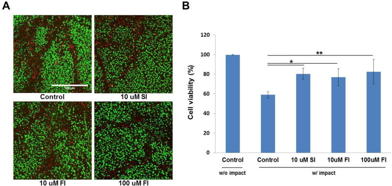
Figure 3. Confocal microscopy and cell viability immediately after impact injury.
(A) Confocal micrographs show live (green) and dead (red) chondrocytes in an impact site in an untreated control explant, and in explants treated with 10μM SFKi and either 10 or 100μM FAKi. Compared to control, fewer dead chondrocytes were observed in SFKs or FAKi treated groups. (B) Statistical analysis revealed that chondrocyte viability was significantly higher in SFKs or FAKi treated explants compared to control. Between two tested concentrations, 100μM FAKi was more effective than 10μM. Asterisk represents statistically significant (*p<0.05, **p<0.01). Bars = 500 μm.

