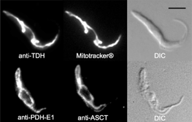
Figure 4.

Immunolocalization of TDH and PDH. Procyclic cells were stained with rabbit anti-TDH (Alexa 488 channel) and MitoTracker® (top panels) or mouse anti-PDH-E1α (Alexa 488 channel) and rabbit anti-ASCT (Alexa 594 channel) (lower panels). Differential interference contrast (DIC) of cells is shown to the right of each panel. Scale bar, 5 μm.
