Figure 6.

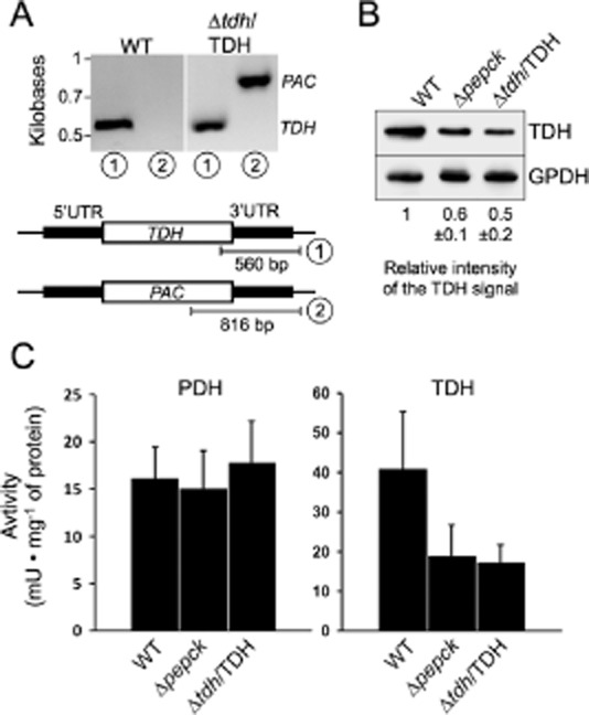
TDH expression and activity are reduced in the Δpepck cell line. Demonstration of the single TDH allele replacement in the Δtdh/TDH cell line is presented in panel A. PCR analysis of genomic DNA isolated from the WT and Δtdh/TDH cell lines was performed with primers based on sequences that flank the 3′UTR fragment used to target depletion of one TDH allele (black boxes) and internal sequences from the TDH gene (PCR product 1) or the PAC (PCR product 2). As expected, PCR amplification using the primer derived from the PAC gene was only observed for the Δtdh/TDH cell line. In panel B, expression of TDH and glycerol 3-phosphate dehydrogenase (GPDH) was analysed by Western blotting with specific immune sera. Ratio between the TDH and GPDH signals, indicated below the blot, represents a mean ± SD of 3 different experimental duplicates, with an arbitrary value of 1 for the parental cells (WT). Panel C shows the PDH (right panel) and TDH (left panel) activities (milliunits mg−1 of protein), normalized with the malic enzyme activity measured in the same samples, i.e. WT, Δpepck and Δtdh/TDH cell lines.
