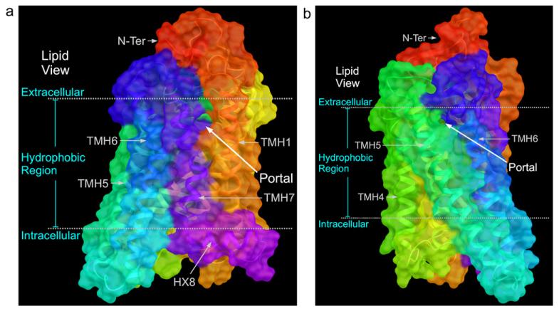
Fig. 5.
The ligand free apoprotein opsin crystal structure (Park et al., 2008) is illustrated here. In (A), the view point is from the lipid bilayer looking towards TMH7 and TMH1. Here one can see that an opening between TMH7 and TMH1 exists. In (B), the view point is from the lipid bilayer looking towards TMH5 and TMH6. Here one can see that an opening between TMH5 and TMH6 also exists The openings illustrated in (A) and (B) have been proposed to be portals such that ligand movement from the lipid bilayer into opsin would be possible (Hildebrand et al., 2009; Schadel et al., 2003).

