
Figure 2.
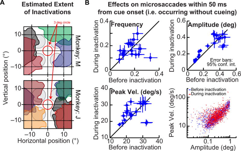
Assessing the spatial extent of inactivations. (A) Each colored region in the figure shows our estimate of the affected region of visual space after a given muscimol injection into the SC. This estimate was obtained based on measured reductions in peak velocities of visually guided saccades (Lovejoy & Krauzlis, 2010; Zenon & Krauzlis, 2012). The shown sessions are from the saccade version of the task, and they were also shown previously in (Lovejoy & Krauzlis, 2010). For reference, the gray circles show the locations of the cue rings in the task of Fig. 1 and the red circle shows a 3-deg radius, indicating that none of our injections extended into the rostral (or foveal) region of the SC. (B) To check for a possible rostral spread of muscimol, which would impair microsaccade generation, we analyzed several microsaccade characteristics before and during SC injections: the fraction of trials containing microsaccades (upper left panel), the amplitude of movements (upper right panel), their peak velocity (lower left panel), and their ‘main sequence’ relationship (lower right panel). In all panels, we performed these analyses for movements occurring within 50 ms from cue onset (i.e. during a baseline, pre-cue fixation interval), and in all panels but the lower right one, each symbol indicates an individual experiment (the lower right panel combines the sessions). Error bars denote 95% confidence intervals.