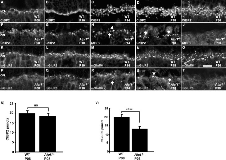
Figure 1.
Reduced expression of mGluR6 prior to photoreceptor loss in Aipl1−/− animals. Immunostaining for CtBP2 (A–J) and mGluR6 (K–T) in WT and Aipl1−/− retina at specified ages. Arrow indicates abnormal staining of CtBP2 (H, I) and mGluR6 (R, S) in the outer plexiform layer of retina from Aipl1−/− animals. Scale bar: 10 μm. Images were obtained using confocal microscopy at 63× magnification with 2× zoom. Average number (±SD) of CtBP2 (U) and mGluR6 (V) puncta in a retinal cross window of 66.8 × 66.8 μm in Aipl1−/− and WT retina at P8. ns, nonsignificant; ****P < 0.0001.

