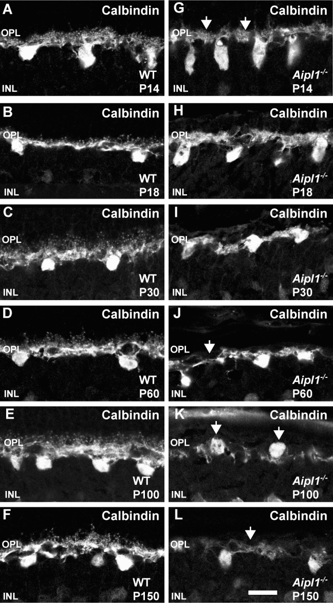
Figure 6.

Altered horizontal cell morphology in the degenerating retina. Calbindin staining reveals changes in the horizontal cell morphology in WT (A–F) and Aipl1−/− (G–L) retina. Arrows point to the loss of puncta from OPL at P14 in retina from animals lacking Aipl1−/− compared to controls (G). (J–L) Arrows indicate thinning of horizontal cell processes and abnormal orientation of cell bodies as animal ages. Scale bar: 20 μm.
