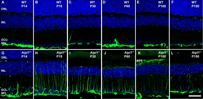
Figure 9.
Activation of Müller glia in the degenerating retina. Müller glial cell morphology and activation were assessed at indicated ages using an antibody against glial fibrillary acidic protein (GFAP; green) in WT (A–F) and Aipl1−/− (G–L) retina. In Aipl1−/− retina, GFAP immunoreactivity is found from nerve fiber layer to the outer retina. DAPI nuclear staining is shown in blue. Scale bar: 20 μm.

