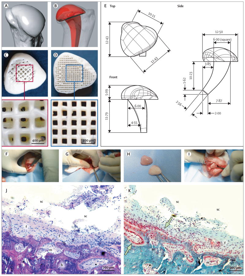
Figure 1. Surgical replacement of synovial joint.
Surface morphology of a rabbit joint was reconstructed (A) to design an anatomically correct bioscaffold (B) with an intramedullary stem. A 200-μm thick shell was designed, along with internal microchannels opening to the synovium cavity (C) and bone marrow (D). PCL-HA was used to fabricate bioscaffolds following computer-aided design (E). The humeral head was excised at its metaphysis junction (F), and an orthopaedic drill used to create an intramedullary tunnel for stem fixation (G). The bioscaffold (H) was implanted by press-fitting (I). In defect-only rabbits, haematoxylin and eosin staining 4 months after surgery (J) showed that little bone had regenerated in the defect; the synovial joint cavity (sc) was visible with fibrous tissue (f) covering bone and marrow (m). Safranin O staining (K) showed scarce chondrocyte-like cells (shown by arrows) in the defect area in the synovial cavity. PCL-HA=poly-ε-caprolactone hydroxyapatite.

