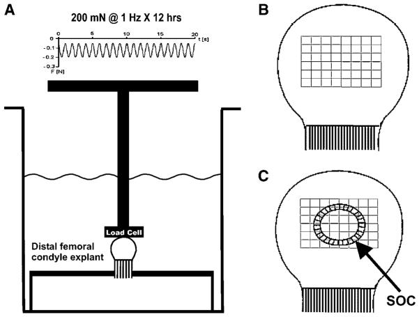
Figure 1.
Schematic diagrams of the neonatal (5-day-old) rabbit distal femoral condyle explant in organ culture with or without exogenous cyclic (intermittent) mechanical loading, as well as methods for computer-assisted histomorphometric quantification. (A) Cyclic mechanical loading at 200 mN and 1 Hz over 12 h was applied to aseptically isolated condyle explants in organ culture. A representative force waveform was shown on the top. At 1 Hz, there was 1 cycle of loading per second. Negative values denoted compressive loading. The bony rudiment of the condyle explant was fixated rigidly. Due to the convex curvature of the articular surface of the condyle explant, both compressive and shear stresses were likely generated. The contralateral condyle explant per rabbit was immersed separately in organ culture medium, but was not subjected to exogenous cyclic loading. (B) Schematic diagram showing an unloaded control specimen with grid blocks (each 900 μm2) constructed in the epiphysis. (C) Schematic diagram showing the formation of the secondary ossification center (SOC) in mechanically loaded condyle explant. The same grid blocks (each 900 μm2) were constructed concentrically from the geometric center of the SOC for computer-assisted histomorphometric analysis as described in the text.

