
Figure 5.
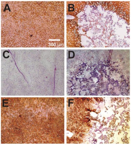
Representative immunolocalization of type II collagen, type X collagen, and decorin in both control (left column) and loaded (right column) neonatal rabbit distal femoral condyle explants. (A) Control explant showing type II collagen expression thoroughly in the chondroepiphysis. (B) The cyclically loaded explant showing little type II collagen within the secondary ossification center (SOC). (C) Control explant showing a general lack of type X collagen expression, which is a chondrocyte hypertrophy marker. (D) The cyclically loaded condyle explant showing sparse type X collagen expression within the SOC. (E) Control explant showing decorin expression thoroughly in the chondroepiphysis. (F) The cyclically loaded explant showing positive decorin expression in chondroepiphysis, but only sparse decorin expression within the secondary ossification center (SOC). [Color scheme can be viewed in the online issue, which is available at http://www.interscience.wiley.com]