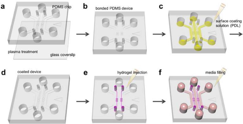
Figure 3.
Procedure of hydrogel incorporating microfluidic assay preparation. (a) Autoclaved PDMS device and coverslip are assembled with plasma treatment (b) to close the microfluidic channels. (c) PDL solution is filled and device is placed in an incubator. (d) After washing and aspiration, coated device is stored in dry oven for 24 hours to render the microfluidic channel surface hydrophobic. (e) Hydrogel is filled into the hydrogel region, and (f) medium is added into the microfluidic channel. Device is ready for cell seeding in incubator.

