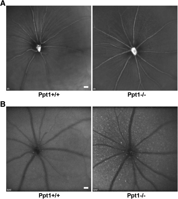
Figure 2.

Funduscopy of 7-month-old Ppt1 +/+ and Ppt1 -/- mice. A: IR fundus images revealed no obvious differences in general retinal appearance of Ppt1 -/- mice in comparison with controls (n = 4 per group). Optic disc and major blood vessels were well recognizable. B: BAF funduscopy demonstrated accumulation of hyperfluorescent profiles in the inner retinal layers of Ppt1 -/- mice in comparison with Ppt1 +/+ mice. Scale bars correspond to 50 μm of subtended retina.
