Figure 8.

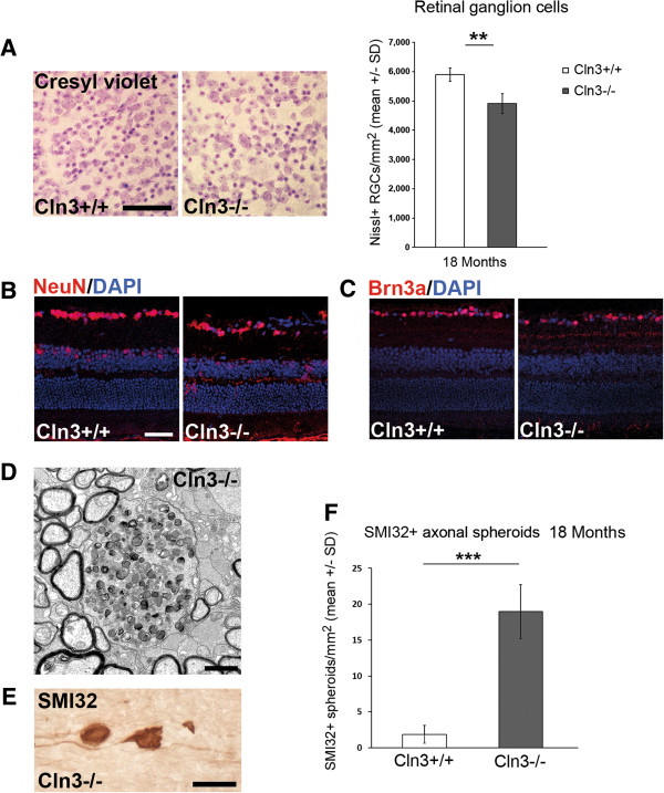
Loss of retinal ganglion cells and axonal perturbation in optic nerves of Cln3 -/- mice. A: Cresyl violet staining of retinal flat mount preparations and quantification of Nissl + retinal ganglion cells in 18-month-old Cln3 +/+ and Cln3 -/- mice (n = 4 per group). Scale bar: 50 μm. B: NeuN and C: Brn3a immunohistochemistry on retinal sections from 18-month-old Cln3 +/+ and Cln3 -/- mice. Scale bar, 30 μm. A loss of NeuN + or Brn3a + retinal ganglion cells in Cln3-/- mice was obvious. D: Representative electron micrograph of optic nerve cross-sections from 18-month-old Cln3-/- mice. Axonal spheroids with accumulation of organelles and dense bodies were frequently observed. E: Immunohistochemistry using antibodies against non-phosphorylated neurofilaments (SMI32; brownish precipitate) in longitudinal optic nerve sections of 18-month-old Cln3 -/- mice visualizes axonal spheroids. Scale bar: 30 μm. F: Quantification revealed that SMI32+ axonal spheroids are barely detectable in optic nerves of Cln3 +/+ mice but accumulate in 18-month-old Cln3 -/- mice (n = 4 per group). Student’s t-test. ***P < 0.001.
