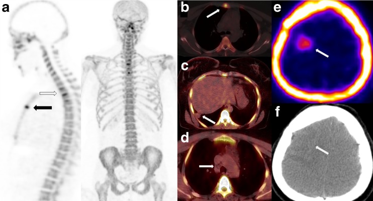
Fig. 2.
Sagittal and coronal MIP images (a) of 18F-NaF PET/CT performed because of severe back pain following the fourth cycle of chemotherapy demonstrate metastases in the thoracic vertebrae (black arrow) and sternum (white arrow). Sternum metastasis is also evident on axial PET/CT images (b). Metastatic pulmonary lesion in the posterolateral segment of the right lung (c) and metastatic right lower paratracheal lymph nodes (d) did not demonstrate any uptake on 18F-NaF PET/CT images. However, the metastatic brain lesion showed 18F-NaF uptake (e) with no visible calcification on the CT counterpart (f)

