
Figure 4.
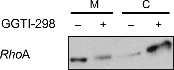
Typical results from subcellular fractionation experiments described in Step 2C. With some proteins, e.g., RhoA, it is notoriously difficult to demonstrate a mobility shift upon PTI treatment. In this case, we recommend preparing cytosolic and membrane fraction, followed by western blotting. The figure (from ref. 79) shows accumulation of RhoA in the cytosol following treatment of CaLu-1 cells with GGTI-298.