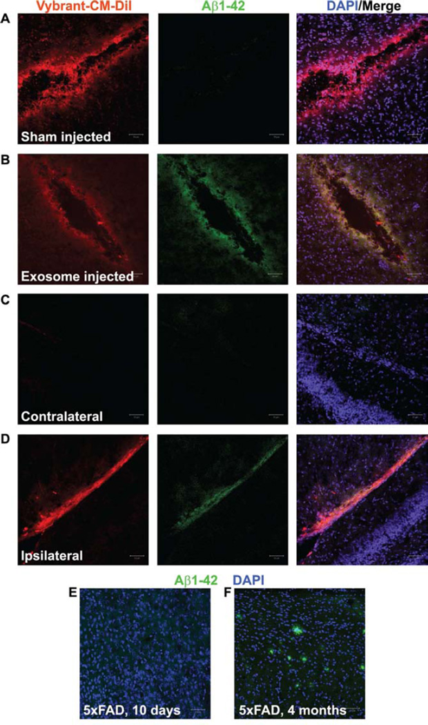
Fig. 2.
Injection of exosomes into 5xFAD mouse brains stimulates aggregation of Aβ1-42. (A-D) Confocal micrographs of 5xFAD mouse brain sections following sham injection (A, control) or injection of astrocyte-derived exosomes (B,D) performed on 10-day old 5xFAD pups that were sacrificed 48 h later. Exosomes were harvested from astrocytes pre-labeled with Vybrant CM-DiI (B,D, red) and resuspended in PBS. Control injections (A) contained 0.01% Vybrant CM-DiI in PBS. All sections were labeled with rabbit anti-Aβ1-42 and AlexaFluor488- anti rabbit IgG (green). Nuclei were labeled with DAPI (blue). Note that sham-injected (A) and the area contralateral (C) to the exosome injection site (D) do not show an accumulation of Aβ1-42. (E-F) Positive and negative controls, respectively, are represented by an adult 5xFAD brain (E) and an uninjected 10-day old 5xFAD pup brain (F). Scale bars = 50 µm.

