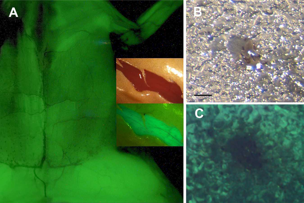
Figure 6. Postmetamorphic expression of XMRF4a transgenes.
(A) GFP fluorescence driven by the 610-bp promoter in skeletal muscle, ventral view, anterior at top, skin removed. This is a composite of two separate exposures of the same animal. Most of the trunk and the left forelimb and proximal hindlimb are shown, but the head is out of the frame. Inset shows reflected-light (upper) and fluorescence (lower) images of a ventral skin incision in the hindlimb of a frog carrying the 1.1-kb promoter. GFP fluorescence in muscle is clearly distinguishable by color from autofluorescence in the skin. (B) Reflected-light image of a 50-µm frozen section of hindlimb muscle from an XMRF4a-1.1-GFP transgenic frog. Note the neurovascular bundle in the center. (C) GFP fluorescence in the same section as in B. Scale bar = 100µm.

