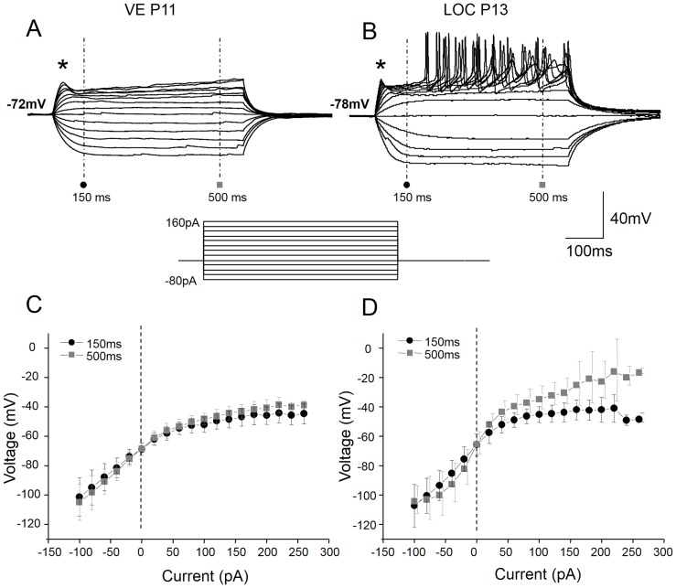
Figure 2. VE neurons have non-linear voltage properties in the depolarizing range.
Voltage responses of VE (A) and LOC (B) neurons to depolarizing current steps of 20 pA (current protocol displayed below the voltage traces) injected from the resting membrane potential. The VE neurons responded with a subtle depolarizing shoulder (asterisk), which failed to trigger action potentials, whilst the LOC neurons show a more pronounced depolarizing shoulder, larger voltage deflections to the same current increments and delayed firing of action potentials upon positive current injection. The voltage-current relationship measured, with respect to the beginning of the recording, both at the onset (150 ms; black circles) and at steady-state (500 ms; grey squares) of the response, displayed strong outward rectification (a decreased slope) in the depolarizing range in the VE neurons, whereas it was near linear in the hyperpolarizing range (C). The LOC neurons also displayed a voltage-dependent non-linearity at the onset of depolarization, but were more linear at steady state voltages (D). The larger variance at steady state are due to the contamination of the spiking activity at the higher current levels in the LOC neurons.

