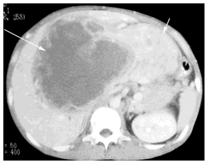
Figure 2.
38-year old male with inflammatory pseudotumor of the liver. IV-contrast enhanced CT scan, portal venous phase, demonstrates a large mass measuring 19.5 by 16.5cm occupying the anterior segments of the right lobe and entire left lobe of the liver (kv 120 mA 180, 150cc of Optiray 380). Most of the cystic/necrotic component is within the anterior and medial segments (long arrow) and the heterogeneous predominantly solid component occupies the lateral segment (short arrow).

