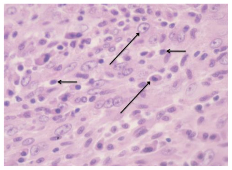
Figure 5.
38-year old male with inflammatory pseudotumor of the liver. High power microscopic view demonstrates a moderately cellular spindle-cell proliferation with heavy inflammatory infiltrate consisting primarily of plasma cells (long arrow) and lymphocytes (short arrow) (40× magnification, hematoxylin and eosin stain).

