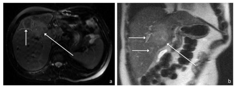
Figure 6.
41-year old male with recurrent inflammatory pseudotumor of the liver. Axial FSE T2-weighted (1.5T, TE 114, TR 2000) (a) and coronal T2-weighted (1.5T, TE 99, TR 1745) (b) MR images in a demonstrate a well-defined hyperintense hepatic mass (long arrow) measuring 5.4 by 3.7cm arising from segment 8 with possible extension into 4A/B. Peripheral biliary ductal dilatation up to 4mm in diameter (short arrows) is also present.

