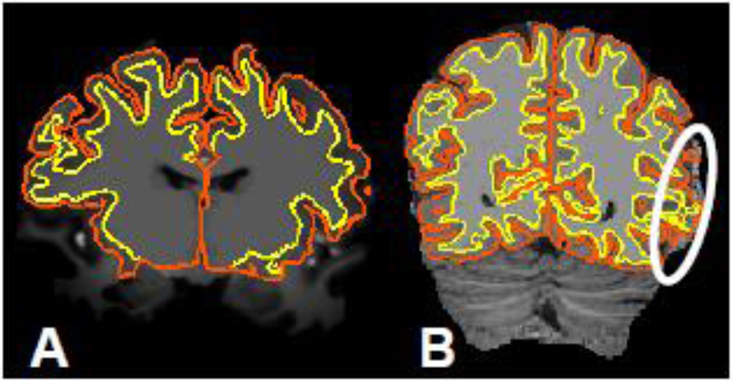
Fig. 1.
(A) Example of an unprocessed 7 T MEMPRAGE image slice with cross-sections of FreeSurfer surface reconstructions generated from this data overlaid. (Yellow contour represents intersection of slice with the white matter surface, while the orange contour represents the pial surface.) (B) Example of a synthetically generated 7 T MP2RAGE “flat image” slice with FreeSurfer surface reconstructions generated using the standard processing stream applied to this data overlaid. The marker indicates a region where the amplified noise in the CSF region causes errors in the cortical GM segmentation, which leads to inaccurate surface reconstructions.

