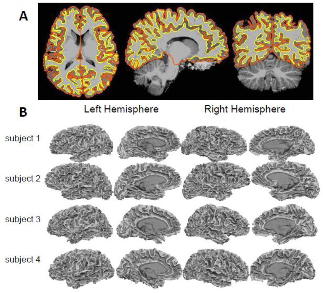
Fig. 4.
Example surface reconstructions from 7 T MP2RAGE data. (A) Masked MP2RAGE flat image with cross-sections of surface reconstructions representing the gray-white and gray-pial boundaries overlaid. (B) Final FreeSurfer white matter surface reconstructions of four subjects, including conventional-bandwidth (240 Hz/pix) and high-bandwidth (975 Hz/pix) examples. (Dark gray indicates sulcal regions and light gray indicates gyral regions.)

