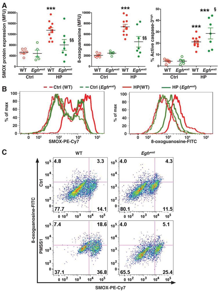
Figure 1.
Levels of SMOX protein, DNA damage, and apoptosis in mice infected with H pylori. C57BL/6 wild-type (WT) and Egfrwa5 mice were infected with PMSS1 for 8 weeks, and gastric epithelial cells were isolated at 2 months post-inoculation. (A) Summary data for levels of SMOX, 8-oxoguanosine, and active caspase-3, measured by flow cytometry. ***P < .001 vs uninfected WT control (Ctrl); §P < .05, §§P < .01 vs infected WT mice. (B) Representative histograms for SMOX and 8-oxoguanosine. (C) Representative dot plots for simultaneous analysis of SMOX and 8-oxoguanosine.

