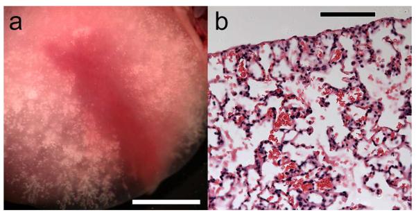
Figure 2.
Photomicrographs of (a) a cranial lung lobe after lavage, which has red color remaining and no residual air retained in the hemorrhage region, and (b) histological image of the residual erythrocytes remaining in the alveolar space after lavage. In (b) the alveolar septa appeared to be intact with no apparent interstitial hemorrhage. Scale bars: (a) 5 mm, (b) 100 μm.

