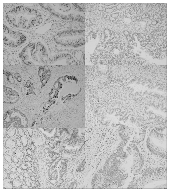
Fig. 3.
Colorectal cancer specimens with typical immunostainings. (Top left) highly positive nuclear p53 immunoreactivity (×200) compared with (top right) a negative tumour. (Middle left) positive nuclear p21 immunoreactivity (×200) compared with a (middle right) a negative sample. (Bottom left) positive MLH1 Immunoreactivity (×200) compared with (bottom right) a negative sample.

