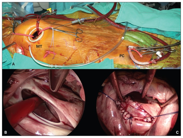
Fig. 1.
(A) Intraoperative photograph demonstrating the 3–4 cm right minithoracotomy (MT) and peripheral cannulation (PC) set-up for minimally invasive, endoscopic atrial septal defect closure. (B) Endoscopic view of a large secundum atrial septal defect with a sump vent going through it. (C) Endoscopic view of the autologous pericardial patch closure of the secundum atrial septal defect.

