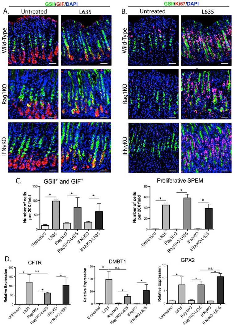
Figure 1. Wild type, Rag1KO and IFNγKO mice develop acute proliferative SPEM after L635-treatment.
A. Immunofluorescence staining for SPEM using gastric intrinsic factor (GIF) in red co-labeled with GSII-lectin (green) and DAPI (blue). GSII-lectin and GIF co-positive cells at the base of the glands were considered SPEM. B. Immunofluorescence staining for proliferation using Ki67 (red) co-labeled with GSII-lectin (green) and DAPI (blue) to determine the presence of SPEM cell proliferation before and after L635 administration in wild type, Rag1KO and IFNγKO mice. Ki67, GIF, and GSII-lectin triple-positive cells were considered proliferative SPEM. C. Quantitation of GSII+ and GIF+ cells (SPEM) and proliferative SPEM cells in wild type, Rag1KO and IFNγKO mice, (*p=0.05). D. qPCR of advanced proliferative SPEM markers. All L635- treated mice showed significant increases in the expression in intestinal transcript markers Cftr, Dmbt1 and Gpx2 (*p=0.05). Scale bars: 50 µm.

