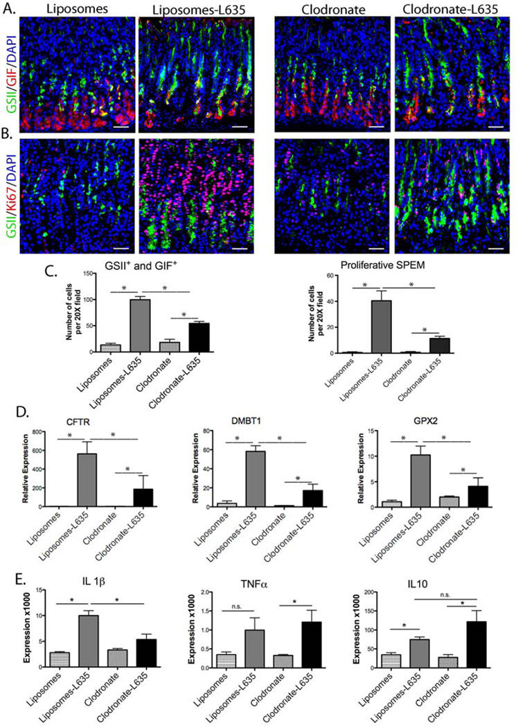
Figure 3. Macrophage depletion inhibits the development of proliferative SPEM following L635 treatment.
A. Immunofluorescence staining of SPEM cells with antibodies against GIF (red) co-labeled with GSII-lectin (green) and DAPI (blue) in mice treated with either control liposomes or clodronate-containing liposomes, with or without L635-treatment. B. Immunofluorescence staining of proliferating SPEM cells with antibodies against Ki67 (red) co-labeled with GSII-lectin (green) and DAPI (blue). C. Quantitation of the number of GSII+ and GIF+ cells (SPEM) and proliferative SPEM cells. Mice treated with both clodronate and L635 had significantly reduced SPEM cell numbers and SPEM cell proliferation (*p=0.05) compared to mice treated with liposomes and L635. D. qPCR showing relative expression of intestinal transcript markers. Clodronate treatment significantly reduced the expression of Cftr, Dmbt1 and Gpx2 in L635-treated mice (*p=0.05). E. qPCR for IL1β, TNFα and IL10 cytokines. Clodronate treatment only significantly reduced IL1β expression (*p=0.05). Scale bars: 50 µm.

