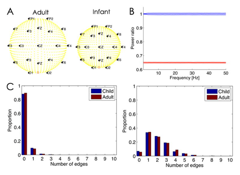
Figure 1. Normalization of power to mitigate impact of skull geometry over development.

A. Illustration of the head geometries in the two model configurations, Adult (left) and Infant (right). The yellow circles denote dipole sources in the cortex, and the black circles scalp electrode locations (with labels). The red square between O1 and O2 denotes the physical reference. B. The ratio of the power spectrum computed in the Adult model configuration divided by the Infant model configuration. The average power is computed for a 2 s interval over all electrodes in each configuration, and then the ratio is determined. The blue line is the power ratio for the normalized spectra. The thick line indicates the mean ratio and the thin lines the 95% confidence intervals over 1000 instantiations of 2 s of pink noise dipole source activity. The sampling frequency is 512 Hz. The mean ratio is near 1, which suggests that the normalization prevents alterations in power due to changes in head geometry. The red line is the power ratio of spectra that have not been normalized; here the mean is smaller because there is less power in the Adult spectra due to the spatial blurring of the thicker skull. C. The number of edges detected in the inferred functional networks depends on the dipole source activity, regardless of head geometry. (Left) When the dipole sources consist of uncorrelated pink noise, both head geometries (Adult and Child, see legend) tend to detect one or fewer edges. (Right) When a subset of dipole sources possess correlated activity, both head geometries detect one or more edges.
