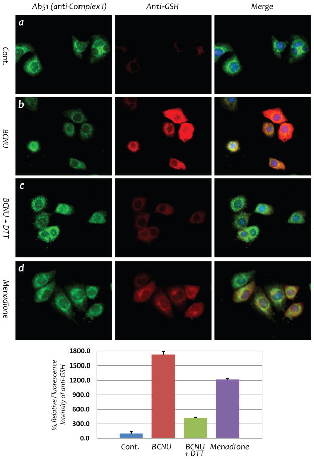
Fig. 6. BCNU enhances S-glutathionylation of Complex I 51 kDa subunit from HL-1 myocyte.
Immunofluorescent detection of S-glutathionylation in HL-1 using an anti-GSH monoclonal antibody was carried out according to the published method (19). Images a and b are control HL-1 myocytes with or without BCNU (25 μM) treatment, demonstrating drastic enhancement of cellular S-glutathionylation by BCNU. Image c is HL-1 treated with BCNU in the presence of DTT (1 mM) in culture, confirming reversible S-glutathionylation induced by BCNU. Image d is the positive control showing increased cellular S-glutathionylation from HL-1 under oxidative stress induced by menadione (40 μM) (19).

