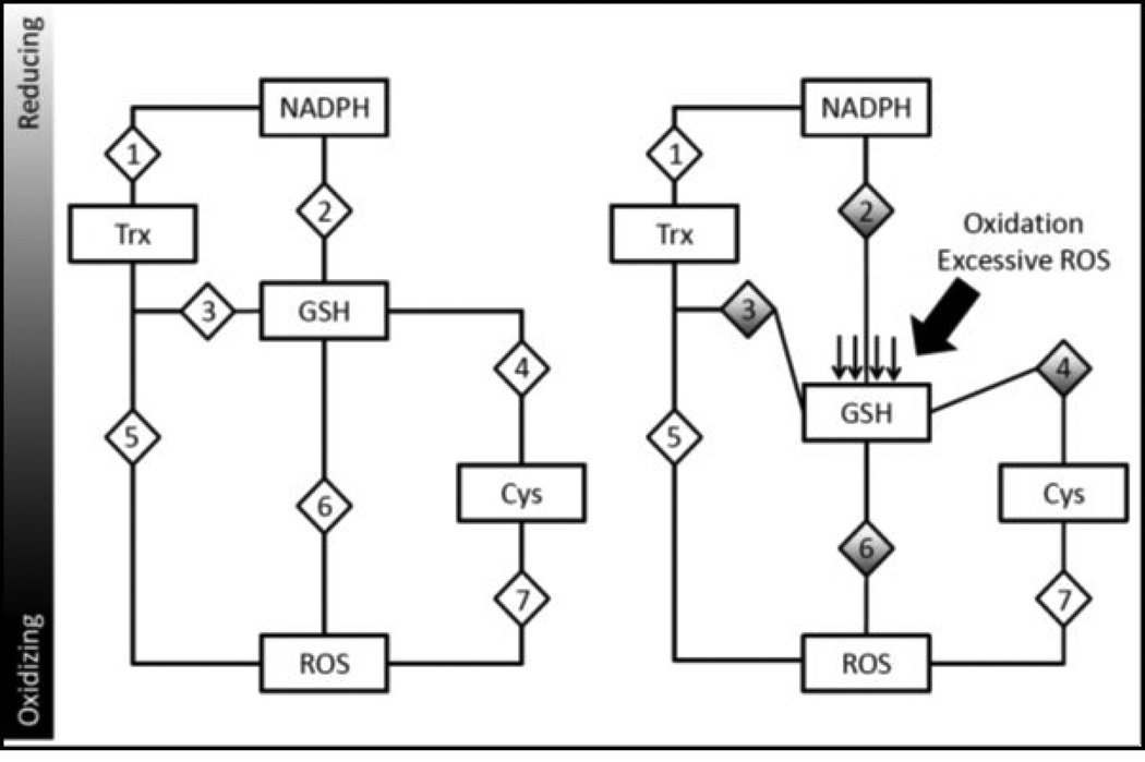
Figure 3.
Redox nodes and circuitry: A mechanism of redox control and dysfunction during oxidative stress. Various redox couples are maintained at various redox states. As these couples are independently regulated, they could serve to control specific subsets of proteins. For example, a hypothetical redox-sensitive protein (diamond labeled ‘3’) may be reduced by thioredoxin but oxidized by GSH. During homeostasis (on left), this protein is maintained in a specific state. However, during periods where GSH becomes oxidized (shifting Eh to a more oxidizing state), hypothetical protein 3 would become increasingly oxidized as the influence of a more oxidizing GSH Eh would cause changes to its redox state and affect its function. Similarly, other proteins (hypothetical proteins 2, 4 and 6) under the regulation of the GSH Eh would also be affected. However, proteins that are not under the control of the GSH Eh would be unaffected, such as proteins 1, 5 and 7. This model of oxi dative stress demonstrates specificity of redox-signaling and provides rationale for ROS-mediated signal transduction.

