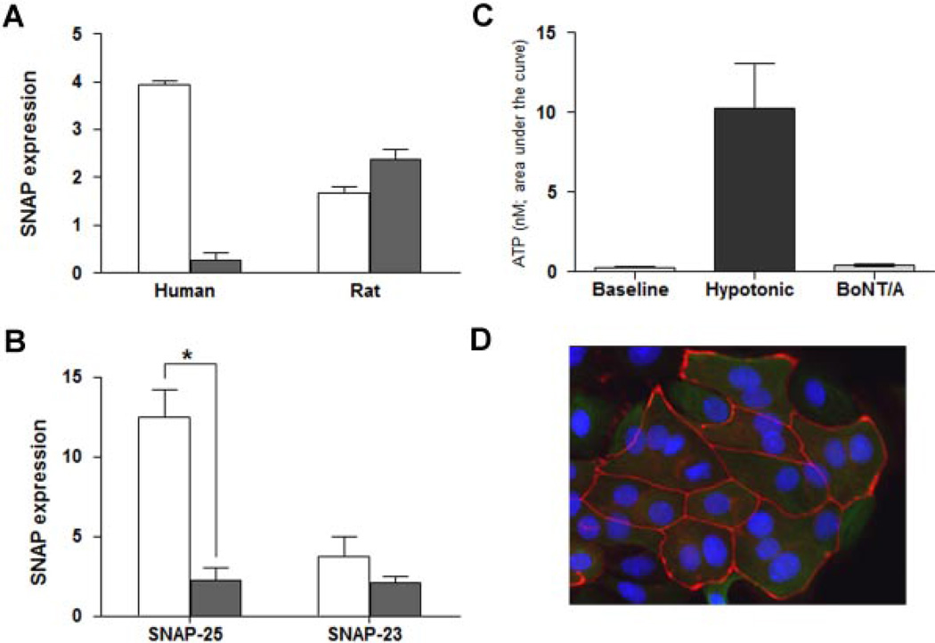
Fig. 2.
The effect of BoNT/A on SNAP-25, SNAP-23 expression and ATP release. A: Summary of Western blot analysis showing protein levels of SNAP 25 (open bars) versus SNAP-23 (solid bars) in both human and rat mucosa. SNAP-25 levels in rat mucosa are less than half of the levels in human mucosa; while SNAP-23 levels in rat mucosa are 10-fold higher as compared to human. Three independent experiments were analyzed and are normalized to beta-actin. B: Levels of SNAP-25 and SNAP-23 protein in control rat urothelial cells (open bars) and urothelial cells treated with BoNT/A (1.5U, for 1–3 hr, solid bars). BoNT/A significantly decreased the expression of SNAP-25 (P<0.05 as compared by unpaired Student’s t-test). C: BoNT/A inhibits hypotonic-evoked release of ATP. Summary graph showing baseline release of ATP, the effect of hypotonic stimuli alone, and hypotonic-evoked release following treatment of cells with BoNT/ A (1.5 U; 1–3 hr). Data are expressed as nM of ATP and were obtained from cultures prepared from at least 6 rats. Experiments were performed on at least n = 6 independent cultures from each animal. P<0.05 as compared by unpaired Student’s t-test. D: Prolonged exposure to BoNT/A does not induce deleterious changes in urothelial cell morphology. Shown is a representative fluorescent image of cultured rat urothelial cells 24 hr following incubation with BoNT/A. The cells were fixed (4% paraformaldehyde) and stained with the junctional marker ZO-1 (shown as red; primary rabbit and secondary antibody Cy3 goat-antirabbit IgG) and counter-stained with the nuclear marker, DAPI (shown as blue).

