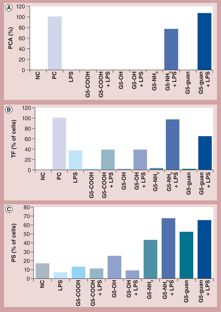
Figure 2. Effects of nanoparticle charge and surface chemistry on procoagulant activity.
Peripheral blood mononuclear cells from healthy donor volunteers were treated with LPS and G5 dendrimers with different surface groups alone or in combination. (A) Induction of PCA by dendrimers at 25 µg/ml and LPS at 1 ng/ml. PCA was calculated as the percentage of coagulation induced by the PC, LPS at 10 µg/ml. Shown is the mean response (n = 4) for one donor. Similar data were obtained with three additional donors (Supplementary Figure S2). (B) Flow cytometry analysis of TF expression on the surface of monocytes after treatment with various dendrimers at 4 µg/ml (G5-COOH; G5-OH; G5-NH2 and G5-guan), LPS at 1 ng/ml or a combination (G5-COOH + LPS; G5-OH + LPS; G5-NH2 + LPS and G5-guan + LPS). Shown is the percentage of positive cells from one donor relative to the PC. Similar data were obtained with two additional donors (Supplementary Figure S2). PC for this test was LPS (10 µg/ml). (C) Flow cytometry analysis of PS presentation on the surface of monocytes after treatment with various dendrimers at 12 µg/ml (G5-COOH; G5-OH; G5-NH2 and G5-guan), LPS at 1 ng/ml or a combination (G5-COOH + LPS; G5-OH + LPS; G5-NH2 + LPS and G5-guan + LPS). Shown is the percentage of positive (i.e., PS presenting) cells. Similar data were obtained with two additional donors (Supplementary Figure S2). PC for this test was 30 min treatment with formalin (not shown).
G5: Generation 5; guan: Guanidine; LPS: Lipopolysaccharide; NC: Negative control; PC: Positive control; PCA: Procoagulant activity; PS: Phosphatidyl serine; TF: Tissue factor.

