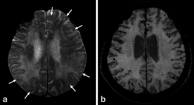
Fig. 1.

Multiple MBs and CAA-related inflammation in a 78-year-old man. In addition to the right dominant diffuse white matter lesions, an axial GRE T2*-weighted image on the 1.5-T imager (a) revealed some cortical-subcortical hypointense foci suggestive of CAA-related MBs (arrows). Of note, more hypointense foci in the posterior dominant distribution were identified on the corresponding PRESTO image (b)
