Fig. 8.

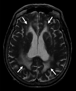
Leukoaraiosis in an 87-year-old woman with pathologically proved CAA and AD. Axial T2-weighted images showed bilateral hyperintensities (arrows), which involved the posterior dominant periventricular and deep white matters

Leukoaraiosis in an 87-year-old woman with pathologically proved CAA and AD. Axial T2-weighted images showed bilateral hyperintensities (arrows), which involved the posterior dominant periventricular and deep white matters