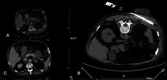
Fig. 3.

a Painful soft tissue mass infiltrating the left T10 posterior rib. b A microwave antenna is percutaneously inserted inside the mass. Due to the proximity to the skin a sterile glove filled with cold water is placed over the skin. c CT axial scan 3 months after the ablation session: Notice the significant size reduction of the mass along with new bone formation around the left T10 posterior rib (similar to heterotopic ossification)
