
Fig. 3.
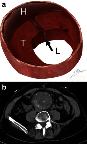
Hyperattenuating crescent sign. a Illustration demonstrates blood (black arrow) dissecting into a mural thrombus (T) from the aortic lumen (L). The resulting intramural haematoma (H) is crescent shaped. b Axial unenhanced CT of a 63-year-old man presenting with abdominal pain and a pulsating mass. A crescent (H) of higher attenuation than the aortic lumen (L) can be seen