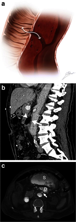
Fig. 6.

Aortoenteric fistula. a Illustration depicts a fistulous tract connecting a bowel loop to an aortic aneurysm. The white double-headed arrow shows communication between the structures allowing bowel gas to infiltrate into the aortic wall and blood to leak into the bowel lumen. b Sagittal enhanced CT image of a 73-year-old man demonstrates an aortoenteric fistula. Intraluminal gas (white arrow) is observed within the AAA, and normal fat planes between the aneurysm and the third portion of the duodenum are lost (white arrowhead). c Axial enhanced CT of an 82-year-old man who presented with massive lower gastrointestinal bleeding and a history of previously repaired AAA. Active contrast extravasation (white arrow) into the third portion of the duodenum (D) and the stomach (S) can be seen in this patient with an aortoenteric fistula
