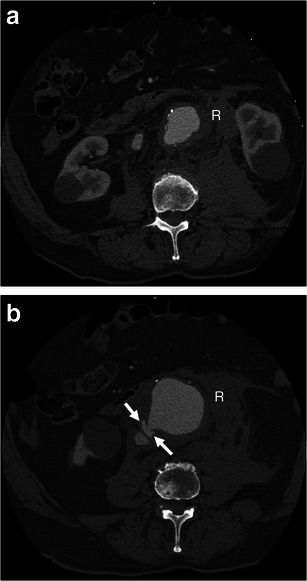
Fig. 7.

Aortocaval fistula. a Axial arterial phase enhanced CT shows simultaneous enhancement of the AAA and IVC in an 83-year-old man with AAA rupture and retroperitoneal haematoma (R). b Axial enhanced CT of the same patient at a lower level demonstrates active contrast extravasation (white arrows) from the aortic aneurysm to the IVC with loss of normal fat planes between the structures. Retroperitoneal haematoma (R) can also be seen
