
Fig. 11.
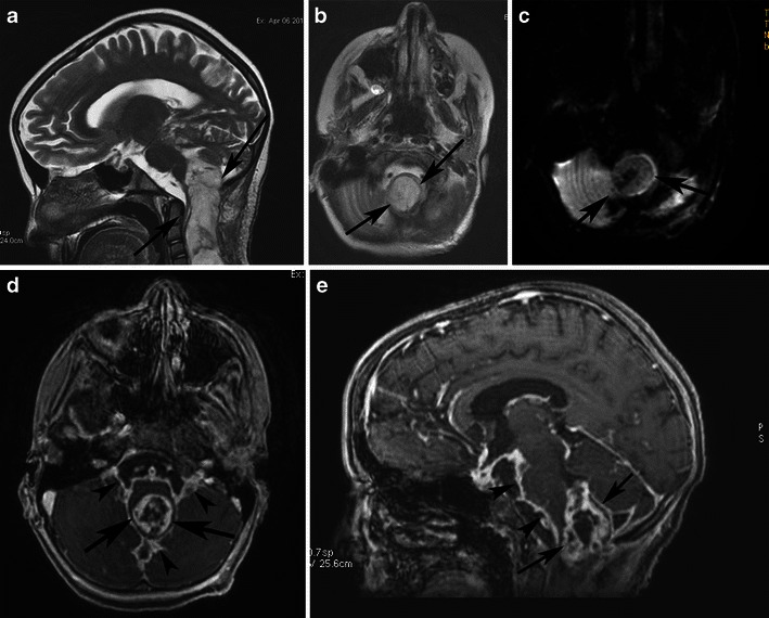
A pilocytic astrocytoma extending to the upper cervical spine with dissemination after partial resection. a Sagittal T2-weighted and b axial T2-weighted images at the level of the foramen magnum show a high-signal cyst-like mass (arrows). c The lesion is hypointense on diffusion-weighted imaging (arrows). Contrast-enhanced axial (d) and sagittal T1-weighted (e) images of the brain show inhomogeneous enhancement of the mass (arrows), as well as diffuse enhancement of the basilar cisternal spaces (arrowheads)