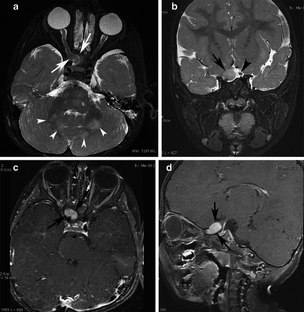
Fig. 14.

A pilocytic astrocytoma of the optic nerve in a 3-year-old girl with NF1. a An axial T2-weighted image reveals characteristic kinking of the enlarged right optic nerve within the intraconal compartment and enlargement (arrow) of the posterior extraconal compartment. Note the characteristic NF1 bright lesions in the cerebellar hemispheres (arrowheads). b A coronal STIR image shows the enlarged right optic nerve (arrow) in comparison with the normal left optic nerve (arrowhead). On these contrast-enhanced axial (c) and sagittal (d) T1-weighted images, intense enhancement of the extraconal component is observed (arrows)
