Figure 6.
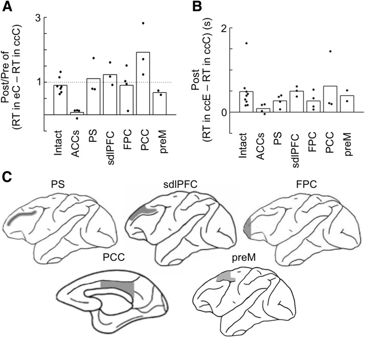
Effects on RT of lesions in various cortical regions. Bilateral lesions were made in ACCs, PS, sdl PFC, FPC, PCC, and preM. A, Postlesion versus prelesion ratio of differences between median RTs in eC trials and median RTs in eciC trials in individual monkeys. The postlesion value was divided by the prelesion value in each monkey, and the ratio was then averaged across monkeys in each group. Bars represent the averaged ratio in each monkey group, and dots indicate the ratios in individual monkeys. The “i” was determined in each monkey so that the difference in %C between after-e and after-eci trials just exceeded 20%. B, Differences between median RTs in ccE trials and median RTs in ccC trials in individual monkeys in postlesion sessions. Bars represent the averaged differences in each monkey group, and dots indicate the differences in individual monkeys. C, Extent of lesions.

