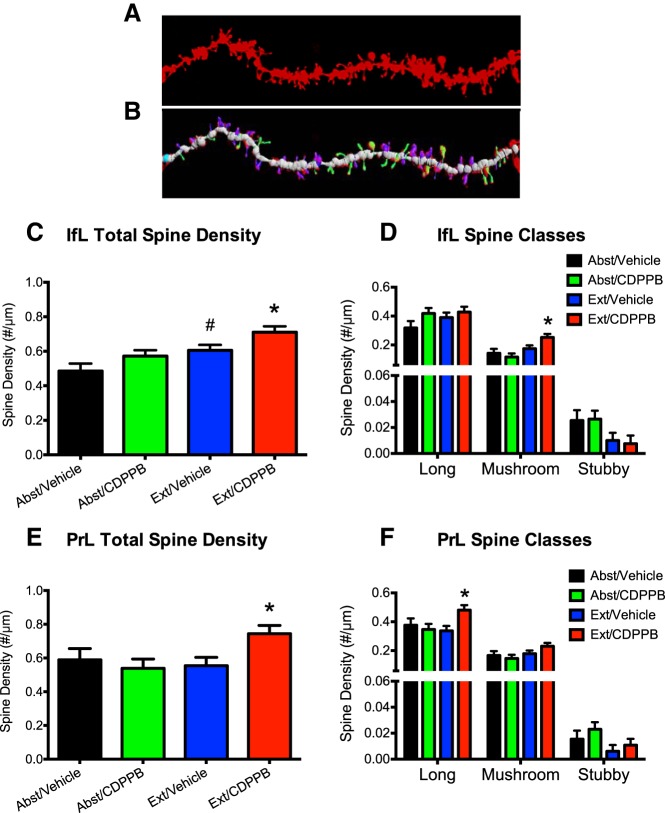
Figure 4.
Facilitated extinction learning is associated with changes in dendritic spines in PrL and IfL cortex. Rats from this experiment were trained to self-administer ethanol using the sucrose-fading paradigm and were killed immediately after the final day of extinction. A, Representative image of a dendrite of a layer 5 pyramidal neuron in the IfL cortex. B, Color-coded image of the dendrite in A showing spine classification based on morphology. C, Extinction training alone was associated with a significant increase in IfL spine density. #p = 0.039. CDPPB treatment further increased spine density during extinction training. *p < 0.038. D, The increase in overall spine density in the IfL cortex resulted from an increase in the density of mushroom shaped spines. *p < 0.03. E, Facilitated extinction learning was also associated with a significant increase in total spine density in the PrL. *p < 0.02. F, The increase in total spine density in the PrL cortex was the result of an increase in the density of immature, long type spines. *p < 0.02. For the Ext/CDPPB (n = 6), Ext/Vehicle (n = 6), Abst/CDPPB (n = 6), and Abst/Vehicle (n = 5) groups, 3–5 neurons were analyzed from the IfL cortex and PrL cortex for each rat and 3–5 dendrites were analyzed and averaged for each neuron.

