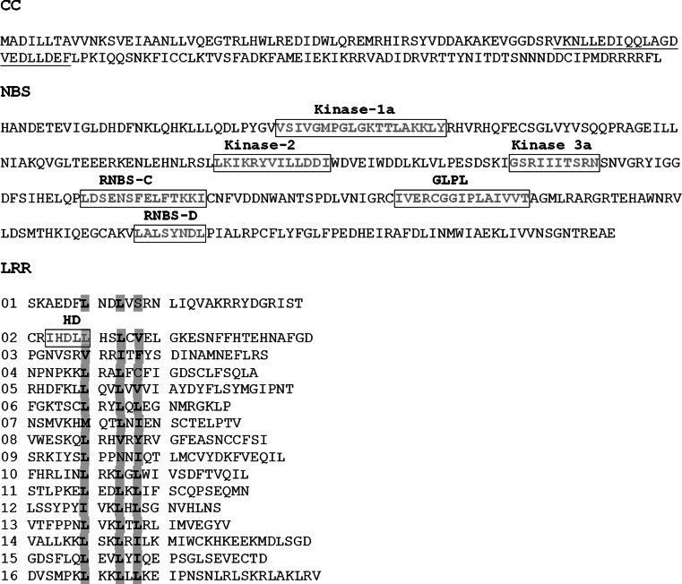
Fig. 6.
The domain structure of the predicted Ph-3 protein. The predicted coiled coil in the CC domain was underlined. Boxes indicate positions of conserved NB-ARC motifs. The 16 imperfect LRRs were aligned according to the consensus sequence xxLxLxx (where L represents leucine or other aliphatic amino acid, and x is any residue)

