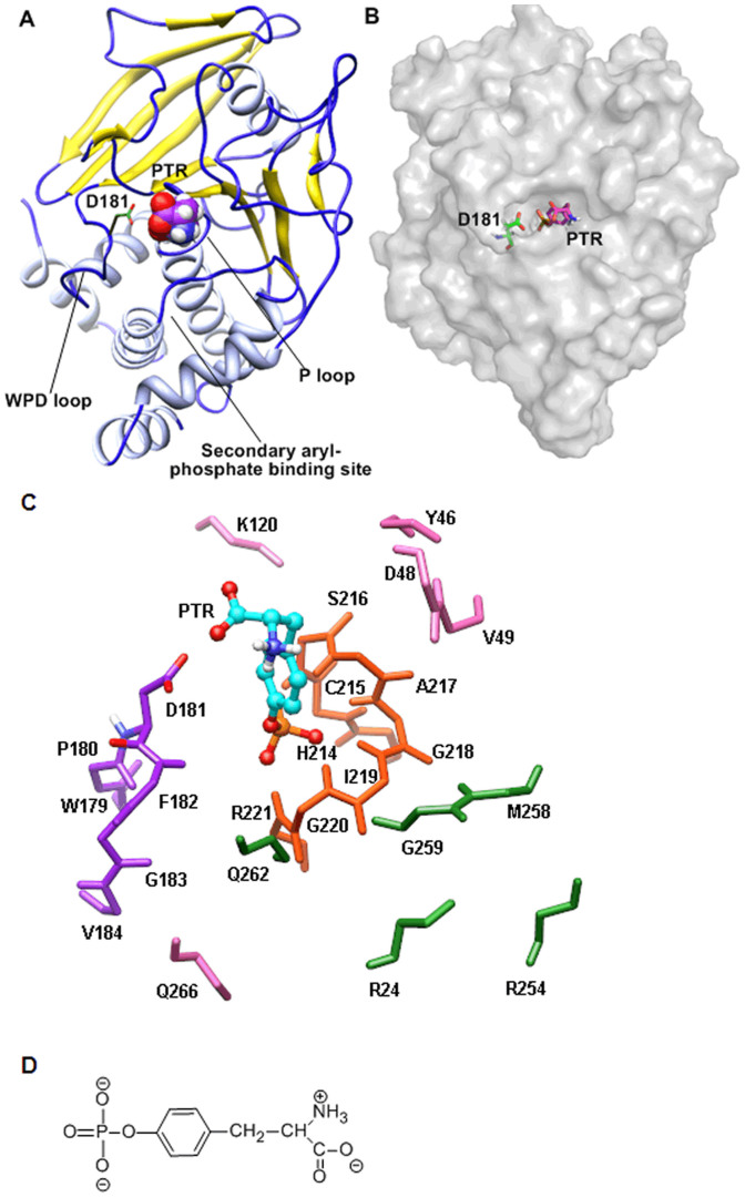
Figure 1. The structure of wild type PTP1B and substrate PTR.
(A) Ribbon structure of PTP1B/substrate complex. (B) The protein surface of PTP1B/substrate complex. (C) The active site of wild type PTP1B. Residues in PTP1B are only shown with backbone atoms except D181. Substrate (PTR) is shown in ball and stick with carbon atoms in cyan. The P loop, WPD loop, secondary aryl-phosphate-binding site and other residues are shown in stick with atoms in yellow, purple, green and pink, respectively. Only polar hydrogen is displayed for clarity. (D) The chemical structure of substrate PTR used in molecular dynamics simulation.

