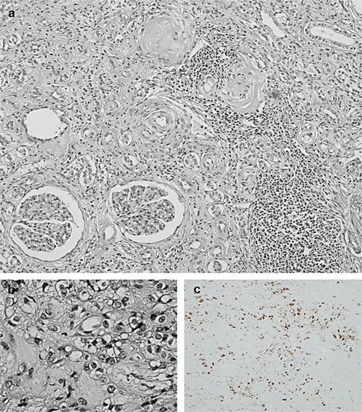
Fig. 1.
a Microscopic findings of the tumor lesion (periodic acid-Schiff stain. ×200). Light micrograph shows a border configuration of the tumor. Glomeruli are surrounded by the tumor lesion. b Microscopic findings of the tumor lesion (periodic acid-Schiff stain. ×400). The tumor is composed of round or spindle-shaped cells containing polygonal nuclei with clear or eosinophilic cytoplasm. Mitotic activity and abnormal mitosis are not observed. c Immunohistochemical staining for renin. Immunohistochemical staining for anti-renin antibody shows a punctate positivity in the cytoplasm and perinuclear area of some tumor cells (immunohistochemistry. ×200).

