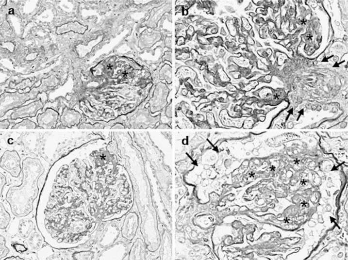
Fig. 3.
Histological characteristics of focal segmental glomerulosclerosis. a Light micrograph shows a segmental area of sclerosis with capillary collapse on the vascular pole (asterisks). b Light micrograph shows a segmental area of sclerosis with capillary collapse on the vascular pole (asterisks) and smooth muscle cell proliferation on the vascular pole (arrows). c Light micrograph shows a segmental area of sclerosis on the urinary pole (asterisk). d Light micrograph shows the collapses of glomerular tufts (asterisks) and cell proliferation on Bowman's capsular epithelium (arrows).

