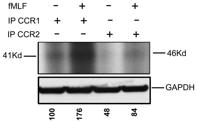
Figure 6. Phosphorylation of CCR1 but not CCR2 after FPR1 activation.

The HR1R2F cells were radiolabeled with 32P-orthophosphate and left untreated or pretreated with 1 μM fMLF. The cell lysates were immunoprecipiated with anti-CCR1 or CCR2 antibodies. The protein samples were dissolved in SDS-loading buffer, and subjected to SDS-PAGE. Phosphorylation of the CCR1 and CCR2 was assessed by autoradiography. The levels of GAPDH were determined by western blot analysis. The normalized absorbance of each of the phosphorylated bands is presented below the GAPDH panel (results are representative of three independent experiments).
